


-
Western blot protocol automation with the ASSIST PLUS pipetting robot and Simple Western™
Foolproof Jess plate set-up with D-ONE
Automated western blotting is a cutting-edge technology revolutionizing traditional protein detection and analysis. Employing robotic systems, microscale separation, and advanced imaging technologies streamlines the labor-intensive and time-consuming process of western blotting. Automating the multiple workflow steps increases efficiency, making it an invaluable tool in various fields of biological research.
Simple Western – developed by ProteinSimple, a Bio-Techne brand – is the only fully automated western blotting solution on the market. The advanced, capillarybased technology enables efficient and accurate high throughput protein separation, detection and quantification, with all assay reagents and samples in 1 plate. Simple Western assays are advancing research and development in many applications, including cancer and immunooncology, cell and gene therapy, regenerative medicine and targeted protein degradation.
The ASSIST PLUS pipetting robot and D-ONE single channel pipetting module effortlessly fill plates specifically designed for Simple Western instruments – like Jess™ or Abby™ – allowing 100 % hands-free time without worrying about tedious liquid handling. INTEGRA protocols automate the Simple Western plate set-up for chemiluminescence detection of single target, multi-target (using RePlex™) or total protein assays. This application note describes fully automated liquid handling of all 3 assay types by performing chemiluminescence detection and total protein analysis in 1 RePlex normalization assay. The results show equivalent performance to manual plate filling, with comparable on-deck sample and reagent stability.
-
Table of contents
Automated western blotting is a cutting-edge technology revolutionizing traditional protein detection and analysis. Employing robotic systems, microscale separation, and advanced imaging technologies streamlines the labor-intensive and time-consuming process of western blotting. Automating the multiple workflow steps increases efficiency, making it an invaluable tool in various fields of biological research.
Simple Western – developed by ProteinSimple, a Bio-Techne brand – is the only fully automated western blotting solution on the market. The advanced, capillarybased technology enables efficient and accurate high throughput protein separation, detection and quantification, with all assay reagents and samples in 1 plate. Simple Western assays are advancing research and development in many applications, including cancer and immunooncology, cell and gene therapy, regenerative medicine and targeted protein degradation.
The ASSIST PLUS pipetting robot and D-ONE single channel pipetting module effortlessly fill plates specifically designed for Simple Western instruments – like Jess™ or Abby™ – allowing 100 % hands-free time without worrying about tedious liquid handling. INTEGRA protocols automate the Simple Western plate set-up for chemiluminescence detection of single target, multi-target (using RePlex™) or total protein assays. This application note describes fully automated liquid handling of all 3 assay types by performing chemiluminescence detection and total protein analysis in 1 RePlex normalization assay. The results show equivalent performance to manual plate filling, with comparable on-deck sample and reagent stability.
Key benefits
provides total protein detection to accurately normalize protein expression data.
Overview: How to fill the Simple Western Jess plate with the ASSIST PLUS
The ASSIST PLUS pipetting robot and D-ONE single channel pipetting module, together with the Simple Western Jess, automate all the liquid handling steps required to analyze 24 samples, providing a complete walk-away solution for western blot protocol automation.
The whole workflow consists of 2 steps (Figure 1):
- Simple Western Jess plate filling with D-ONE and the ASSIST PLUS
- Protein analysis with the Simple Western
This application note provides Jess plate filling protocols for chemiluminescence detection of single targets, multiple targets (using RePlex) and total protein analysis of prepared samples and reagents for reliable downstream protein analysis using Simple Western Technology.
Downloads: App note and protocols for western blot protocol automation with the ASSIST PLUS pipetting robot and Simple Western™
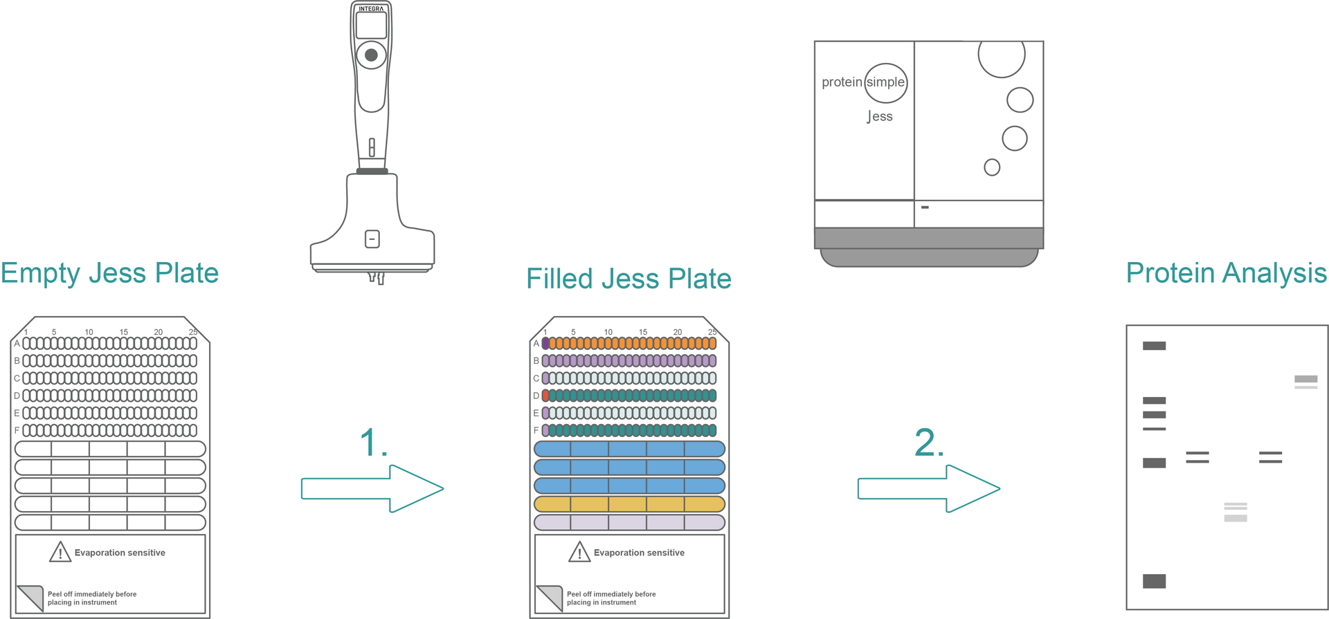
Plate set-up for RePlex chemiluminescence western blot protocol automation
Experimental set-up:
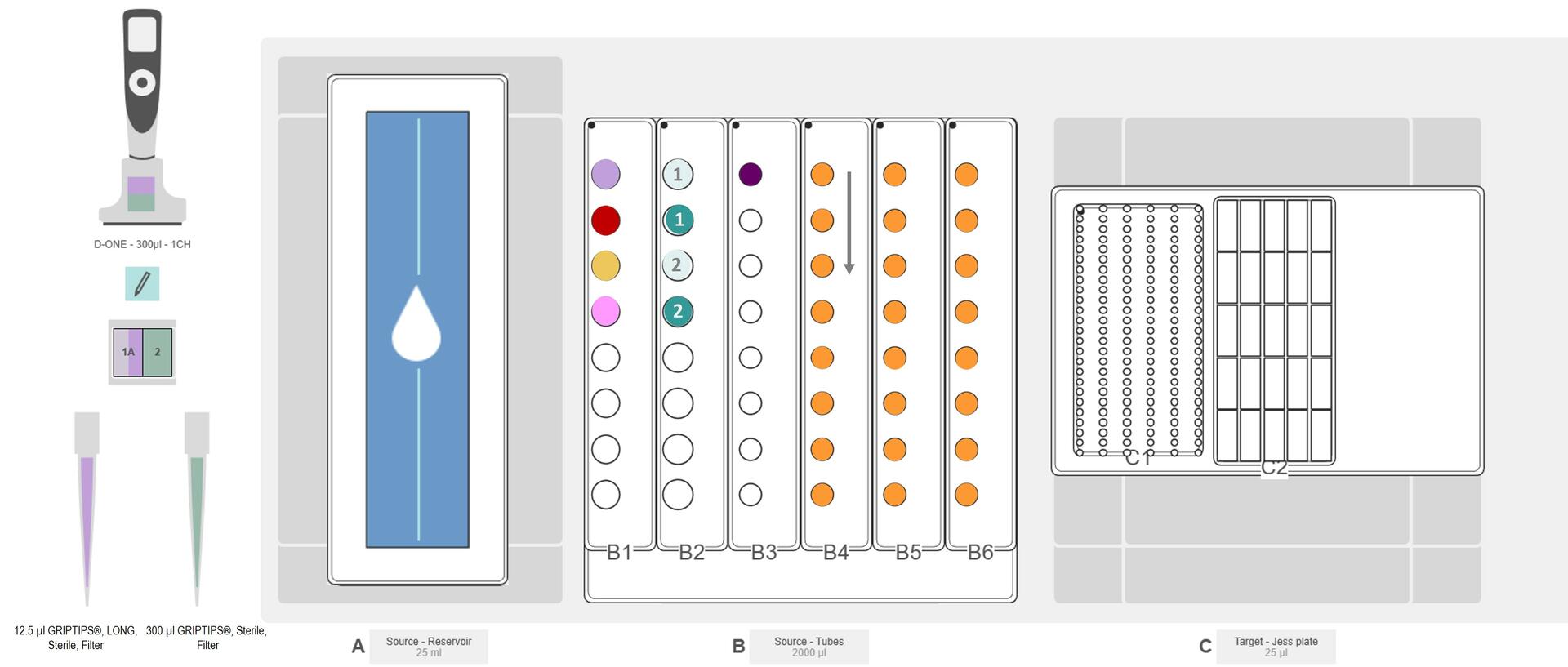
Deck Position A: Wash buffer (blue)
Deck Position B: B1 – antibody diluent (lavender), streptavidin-HRP (red), luminol-peroxide mix (yellow), RePlex mix (pink); B2 – primary antibody probe 1 (light green-1), secondary antibody probe 1 (green-1), primary antibody probe 2 (light green-2), secondary antibody probe 2 (green-2); B3 – biotinylated ladder (violet); B4-B6 - prepared samples (orange, arrow indicates processing direction)
RePlex plate set-up
Jess plate set-up for RePlex
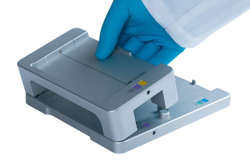
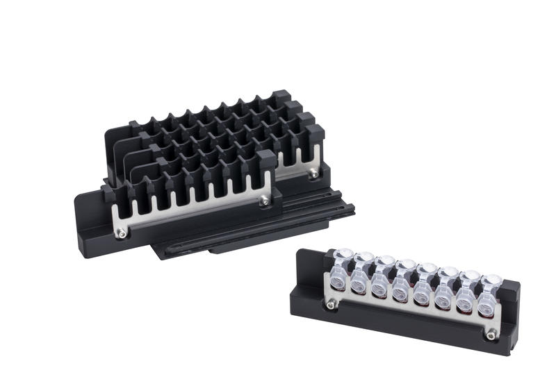
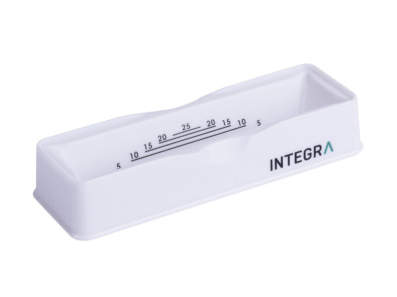
Place a 25 ml reservoir on deck Position A and fill it with at least 8 ml of wash buffer. Place the INTEGRA tube rack on Position B with the specific reagent and sample tubes, as indicated in the experimental set-up. As shown in Figure 2, the Jess plate is in landscape orientation on Position C. LLD enables the use of different aliquot sizes, and the D-ONE informs the operator if the liquid volume is insufficient.
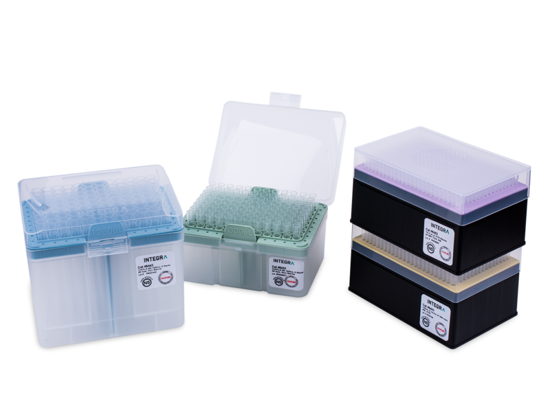
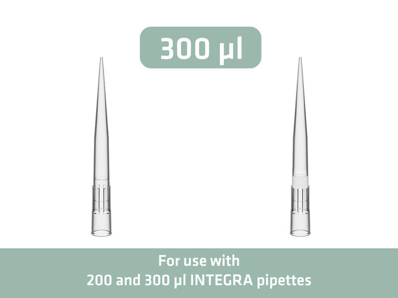
Equip the ASSIST PLUS with the 0.5-300 μl D-ONE, and run the VIALAB program 'Jess_plate_setup_RePlex'. The ASSIST PLUS and D-ONE with 12.5 μl long, sterile, filter GRIPTIPS transfer 10 μl of antibody diluent from a 2 ml tube in Position B1 (Figure 2, lavender) to wells B1-B25, C1, E1 and F1 of the Jess plate (Figure 3) in Position C. A 1 μl pre- and post-dispense guarantees precise pipetting while preventing bubble creation during dispensing. By automatically changing GRIPTIPS between different reagents or samples, the D-ONE transfers 10 μl of primary antibody probe 1 from a 1.5 ml microcentrifuge tube in B2 (Figure 2, light green-1) to wells C2-C25 (Figure 3) of the Jess plate in Position C (Figure 4a). The D-ONE transfers 10 μl of streptavidin-HRP/NIR from the second 2 ml screw cap vial in B1 (Figure 2, red) to well D1 (Figure 3). Wells D2 to D25 (Figure 3) are filled with 10 μl secondary antibody probe 1 from a 1.5 ml microcentrifuge tube in B2 (Figure 2, green-1). 10 μl primary antibody probe 2 is transferred from Position B2 (Figure 2, light green-2) to wells E2 to E25 (Figure 3), and 10 μl secondary antibody probe 2 is transferred from B2 (Figure 2, green-2) to wells F2 to F25 (Figure 3).
Using 300 μl sterile, filter GRIPTIPS, the D-ONE transfers 500 μl of wash buffer (Figure 2, blue) from the 25 ml reservoir on Position A to the Jess plate compartments in 2 steps (Figure 4b), as indicated in Figure 3. A slow speed (5) prevents bubble creation during buffer dispensing. Afterwards, 170 μl of luminol-peroxide mix is transferred from the 2 ml tube in B1 (Figure 5, yellow), and 300 μl of RePlex reagent mix from another 2 ml tube in B1 (Figure 5, pink) to the compartments indicated in Figure 3. The D-ONE then transfers the RePlex reagent mix in 2 steps, with a pre- and post-dispense of 10 μl to prevent bubble creation during dispensing.
5 μl of biotinylated ladder is transferred from a 0.5 ml microcentrifuge tube in Position B3 (Figure 2, violet) to well A1 (Figure 3). As indicated by the arrow in Figure 2, the D-ONE transfers 3 μl of prepared sample (Figure 2, orange) from each 0.5 ml microcentrifuge tube in Positions B4 to B6 (Figure 2, orange) to wells A2-A25 (Figure 3). Fast dispensing (speed 10) increases the accuracy for small volumes. The pipette then instructs the user to centrifuge the plate for 5 minutes at 2500 rpm, before proceeding with Simple Western Jess protein separation and immunodetection.
Tips:
- A prompt can be included in the protocol before starting the sample transfer to instruct the user to prepare the samples during reagent transfer.
- VIALAB's simplified programming allows plate set-up to be easily adjusted to perform western blot normalization by replacing the second chemiluminescence detection with a total protein analysis, as indicated in the kit insert.
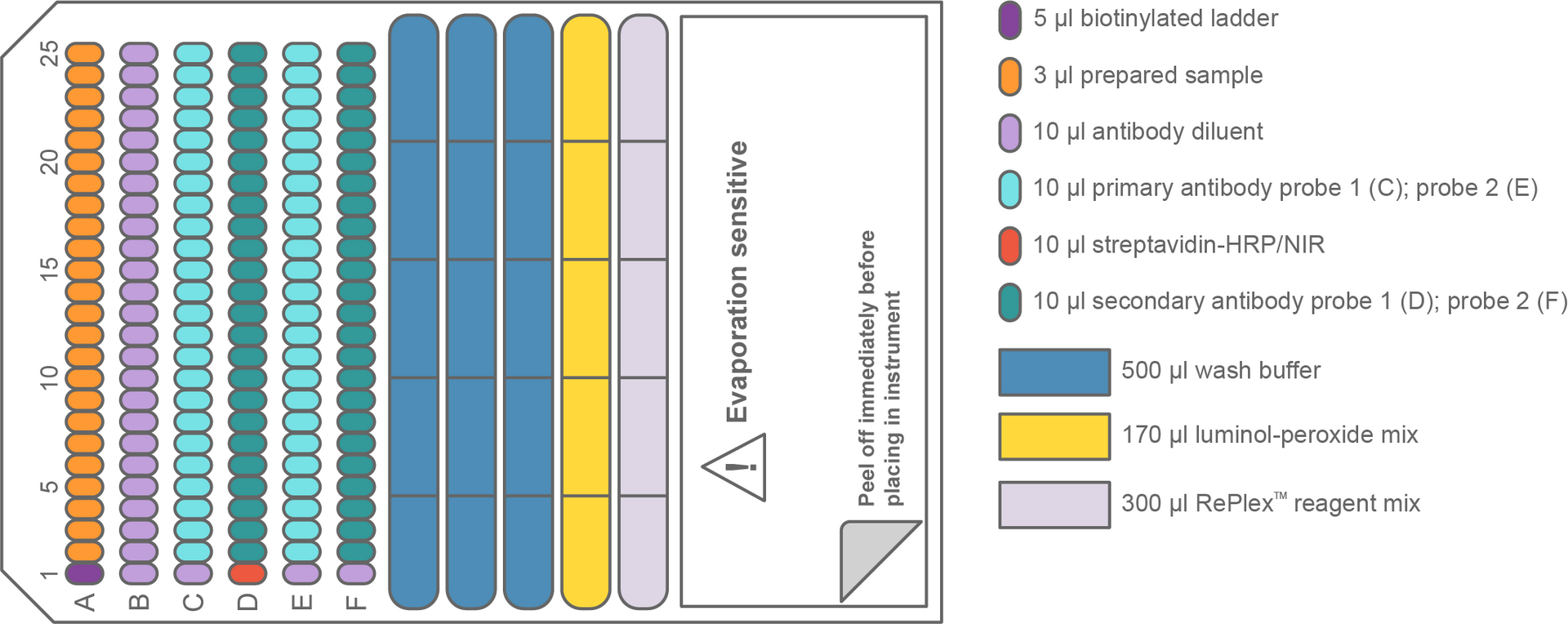
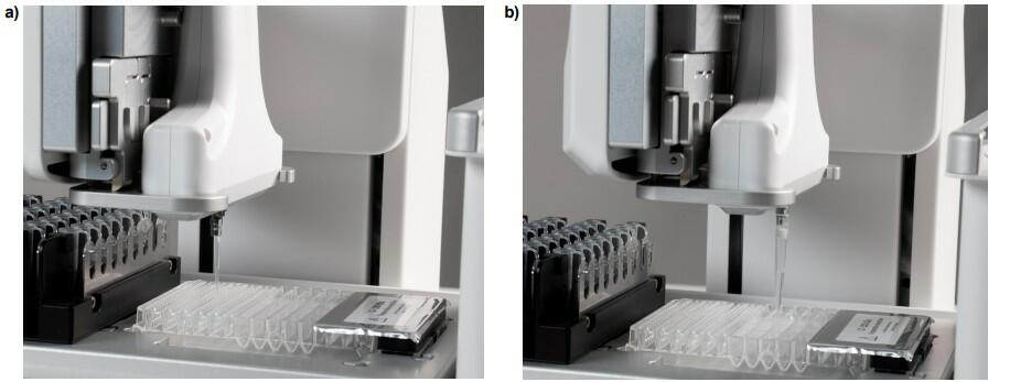
Plate set-up for chemiluminescence western blot protocol automation
Experimental set-up:
Deck Position A: Wash buffer (blue)
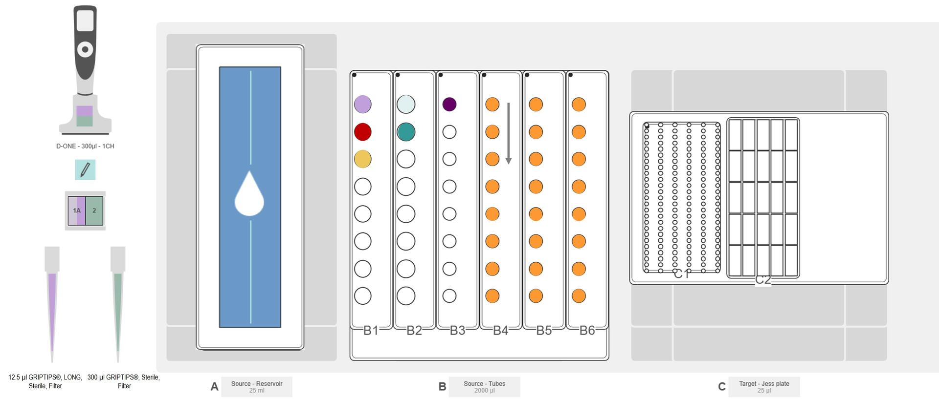
Deck Position B: B1 – antibody diluent (lavender), streptavidin-HRP (red), luminol peroxide mix (yellow); B2 – primary antibody (light green), secondary conjugate (green); B3 – biotinylated ladder (violet); B4-B6 – prepared samples (orange, arrow indicates processing direction)
Chemiluminescence plate set-up
Jess plate set-up for chemiluminescence western blot
Prepare a similar deck set-up to the RePlex western blot protocol, but without the RePlex reagent mix or the primary and secondary antibodies for probe 2 (Figure 5).
Select and run the VIALAB program 'Jess_plate_setup_chemiluminescence'. Following a similar procedure as for RePlex chemiluminescence detection, the D-ONE starts by transferring 10 μl of antibody diluent (Figure 5, lavender) to wells B1-B25 and C1 of the Jess plate (Figure 6) in Position C. 10 μl of primary antibody (Figure 5, light green) is then transferred to wells C2-C25, 10 μl of streptavidin-HRP/NIR (Figure 5, red) to well D1, 10 μl of secondary conjugate (Figure 5, green) to wells D2 to D25, and 15 μl of luminol-peroxide mix to all wells of row E, as indicated in Figure 6.
The D-ONE follows this with transfer of the wash buffer (Figure 5, blue), biotinylated ladder (Figure 5, violet) and samples (Figure 5, orange), as described for RePlex chemiluminescence detection, and instructs the operator to centrifuge the plate before proceeding with the automated western blot.
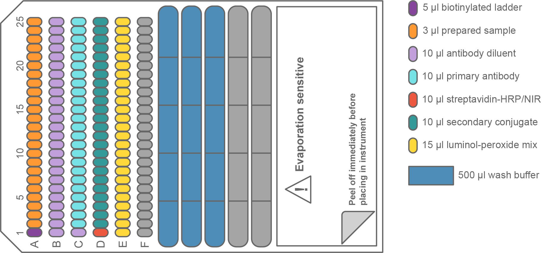
Plate set-up for total protein western blot protocol automation
Experimental set-up:
Deck Position A: Wash buffer (blue)
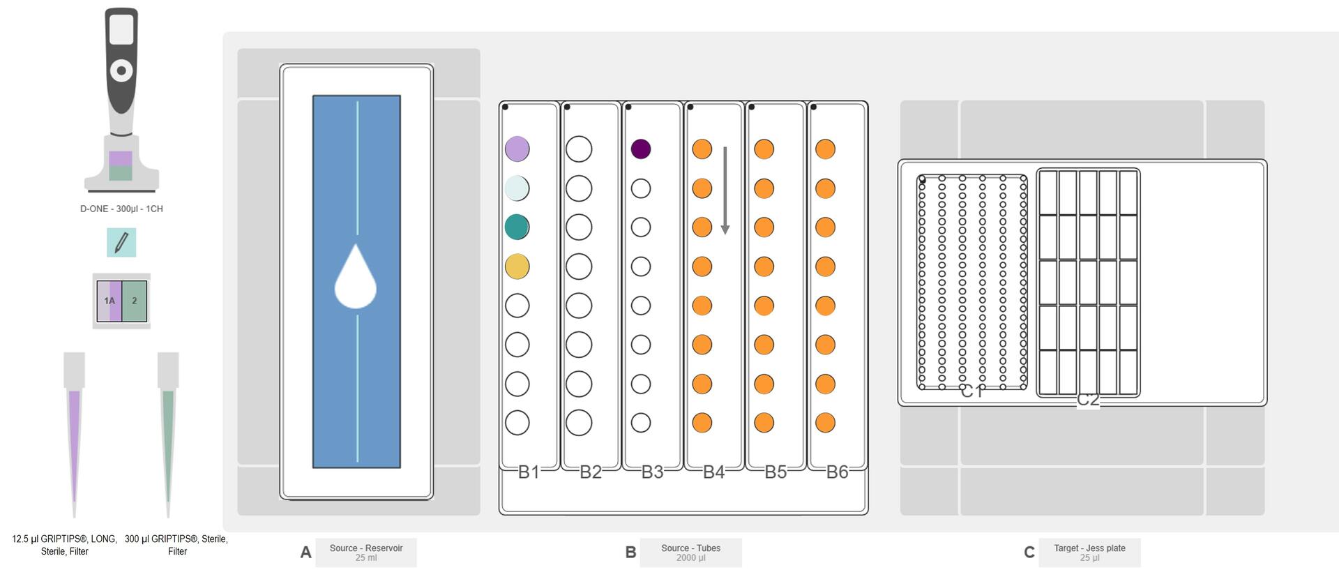
Deck Position B: B1 – antibody diluent (lavender), total protein labeling reagent (light green), total protein streptavidin-HRP (green), luminol-peroxide mix (yellow); B3 – biotinylated ladder (violet); B4-B6 – prepared samples (orange, arrow indicates processing direction)
Total protein plate set-up
Jess plate set-up for total protein analysis
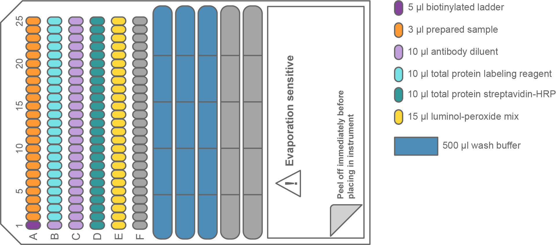
Set up the ASSIST PLUS deck in a similar way to the chemiluminescence detection protocol, but with the reagents for total protein analysis (Figure 7).
Select and run the VIALAB program 'Jess_plate_setup_total_protein'. The D-ONE automatically selects GRIPTIPS, and transfers 10 μl of antibody diluent (Figure 7, lavender) into wells B1 and C1 to C25, 8 μl of total protein streptavidin-HRP (Figure 7, green) to row D, 15 μl of luminol-peroxide mix (Figure 7, yellow) to row E, and wash buffer from the reservoir to the compartment of the Jess plate, as indicated in Figure 8. Similar to the western blot protocol for chemiluminescence detection, 5 μl of biotinylated ladder (Figure 7, violet) and 3 μl of each sample (Figure 7, orange) are also transferred to the Jess plate in Position C. The run is completed by the transfer of 10 μl of total protein labeling reagent (Figure 7, light green) into wells B2 to B25, as shown in Figure 8. After finishing the liquid transfers, the instrument instructs the operator to centrifuge the plate, as indicated in the kit.
Methods and results
Western blotting can be very time consuming and prone to errors. This application note demonstrates a full, walk-away solution for reliable and high throughput automated western blotting, combining INTEGRA's ASSIST PLUS pipetting robot and D-ONE single channel pipetting module with ProteinSimple's Simple Western Jess.
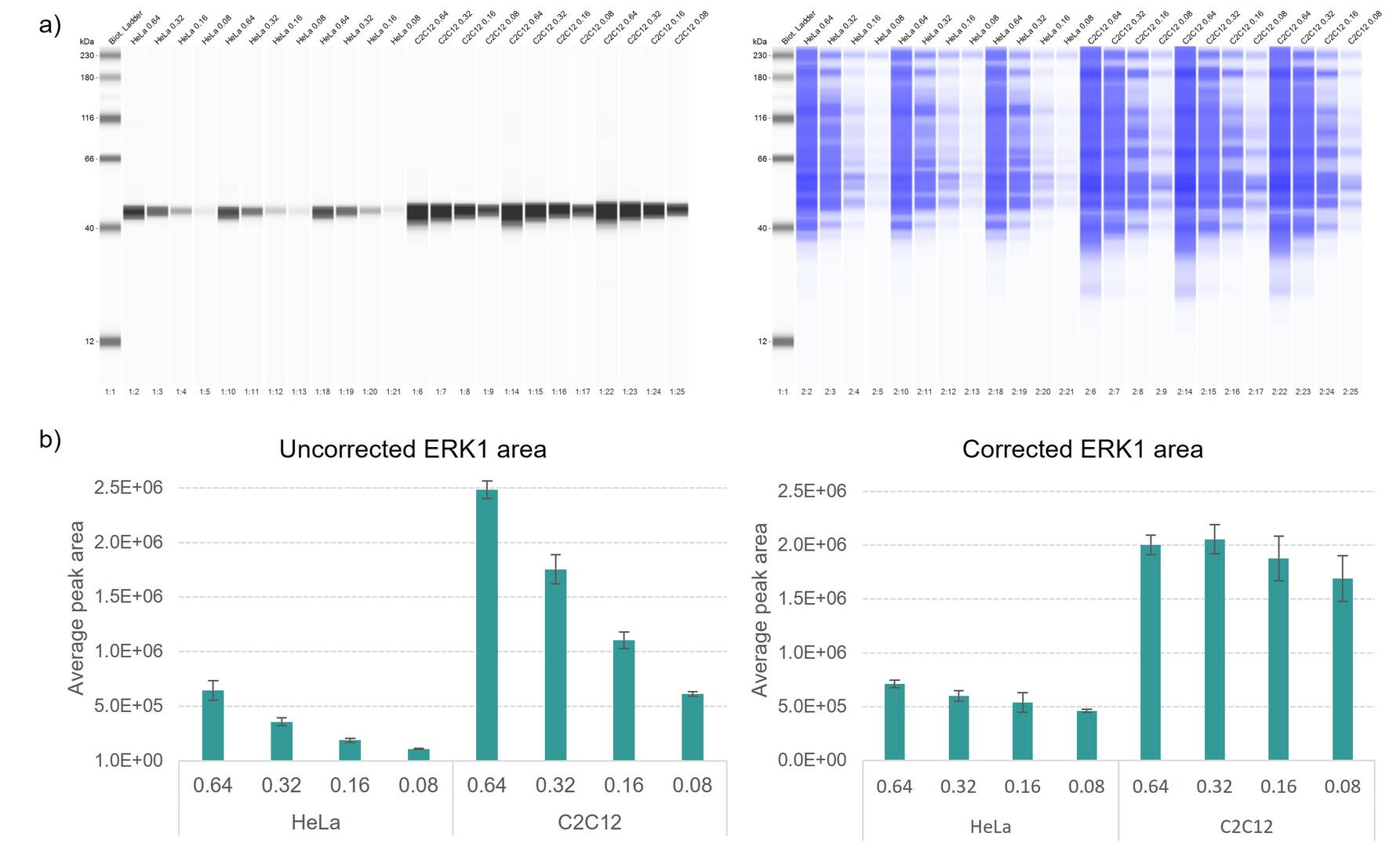
With RePlex, chemiluminescence and total protein detection were combined in a single assay to prove accurate liquid handling for all reagents when setting up plates with the automated protocols. Reagents, HeLa and C2C12 lysates were prepared according to the protocol in the product insert, with lysate dilutions of 0.64, 0.32, 0.16 and 0.08 mg/ml, together with ready-to-use ERK1 primary and secondary antibodies. Each HeLa or C2C12 dilution was prepared in single tube triplicates as individual samples during plate filling.
Figure 9 shows fully automated western blot normalization of HeLa lysates (lanes 2-13), and C2C12 (lanes 14-25), in triplicate. After ERK1 detection (Figure 9a; left), the primary and secondary antibodies were removed with the RePlex reagent mix, to re-stain samples for total protein analysis (Figure 9a; right). The data sets generated were automatically analyzed, using the Simple Western software tool to visualize the uncorrected (Figure 9b; left) and corrected (Figure 9b; right) ERK1 peak target areas. All 3 replicates of each lysate showed great reproducibility while successfully normalizing ERK1 protein levels in 4 different concentrations, proving accurate liquid handling and confirming on-deck reagent/sample stability during plate set-up.
Furthermore, simple fluorescence-based detection was carried out to prove the equivalence in performance between automated and manual plate filling, by removing the luminol-peroxide mix from the VIALAB protocol for chemiluminescence detection (Page 6).
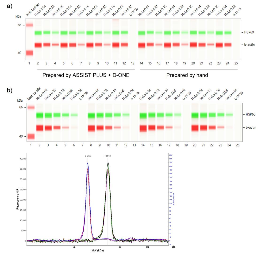
Again, reagents and HeLa lysates were prepared according to the protocol in the product insert, with sample dilutions of 0.64, 0.32, 0.16, 0.08, 0.04 and 0.00 mg/ml (0.1x sample buffer). The primary antibody was prepared by diluting 15 μl of HSP60 and 6 μl of ß-actin in 279 μl of milk-free antibody diluent. The secondary antibody was prepared by diluting 15 μl of anti-rabbit IR and 15 μl of anti-mouse NIR in 270 μl of milk-free antibody diluent.
Figure 10a illustrates the results of automated and manual Jess plate set-up (lane view). Wells 1-13 of rows A to D, as well as the wash buffer compartments, were filled using the D-ONE and ASSIST PLUS. Wells 14-25 of rows A to D were filled manually using a single channel pipette. Both filling methods produced reliable fluorescence data when processing the first 3 dilutions of the HeLa lysates in quadruplicate (0.64 and 0.32 mg/ml) and triplicate (0.16 mg/ml), with CVs within the instrument’s specifications.
Figure 10b illustrates the results when performing fluorescent detection of all 6 HeLa dilutions, by preparing each dilution in 4 single tube replicates on the ASSIST PLUS deck (lane view and electropherogram showing 0.64 mg/ml replicates as representation). Again, CVs met the instrument specifications while showing high detection sensitivity for low abundance targets when automating plate filling with the D-ONE and ASSIST PLUS.
Remarks
- Labware: The simple labware creation tool in the VIALAB library makes the integration of special plates easier than ever.
- VIALAB software: VIALAB programs can be easily adapted to your specific pipette, labware and protocols.
- Partial plates: Programs can be adapted at any time to a different number of samples, giving laboratories total flexibility to meet current and future demands.
Conclusion
Ask our expert. Leave a comment!
Write us if you have any questions regarding the application note or one of our instruments.
Any questions? I'm happy to help!
Instruments and accessories
ASSIST PLUS, Pipetting Robot
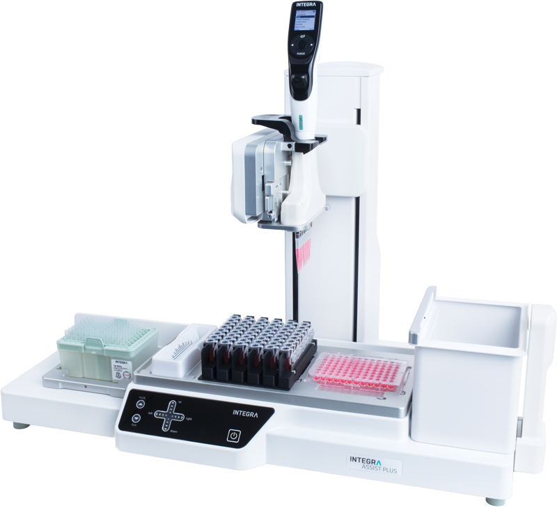
INTEGRA has developed the ASSIST PLUS pipetting robot to streamline routine pipetting tasks at an affordable price. Using INTEGRA electronic multichannel pipettes, the system:
- automates pipetting tasks,
- eliminates physical strain and
- ensures superior reproducibility and
- error free pipetting.
Part No. 4505
Downloads
Download App Note as PDF
DownloadWestern blot protocol automation with the ASSIST PLUS pipetting robot and Simple Western™
Customer’s voice
From ddPCR, to ELISAs and sample normalization for HPLC, CE-SDS and cell based assay useage, we use the ASSIST in our lab daily. We have 6 across our three labs. They are always reliable, accurate, easy to use, and have a very wide range of applications. In addition to freeing up analysts to perform other work, they have decreased assay variation across multiple assays and users in our labs, for more reliable results with more impactful data! The team at INTEGRA is always willing to help with programming, and is very good at listening to feedback in terms of new accessories and software updates. We are very happy with them!
Related products

Applications
Show all-
End-to-end Twist library preparation protocol for NGS workflow automation
-
A simple and streamlined workflow for membrane protein isolation
-
Streamlining single-cell sequencing with the Flex Apex workflow using automated liquid handling solutions
-
Optimizing DNA fragmentation and NGS library preparation with automated liquid handling
Stories
Show all-
 · Company News
Streamlining CRISPR workflows, from editing to analysis
-
 · Customer Testimonial
Enhancing the consistency of ag-tech R&D processes with the ASSIST PLUS pipetting robot
-
 · Product News
Magnetic beads represent a modern approach to gDNA isolation
-
 · Product News
Magnetic beads for NGS size selection and PCR purification