From RNA to results: the essential guide to RT-PCR
Written by Anina Werner
24. April 2025
Reverse transcription-polymerase chain reaction (RT-PCR) – not to be confused with real-time PCR (qPCR) – is a sensitive and specific molecular biology technique that is used to detect and analyze RNA. It enables researchers and clinicians to amplify target RNA sequences, for example, messenger RNA (mRNA) for gene expression studies, or RNA virus genomes for the detection of diseases like COVID-19.
This article explains how RT-PCR works – covering the key steps from RNA extraction to detection – and explores the differences between qualitative RT-PCR and quantitative RT-qPCR. It also compares 1-step and 2-step RT-PCR, helping researchers to determine which method best suits their needs, and provides examples of applications in fields like medicine, forensics and agriculture.
Table of contents
How RT-PCR works
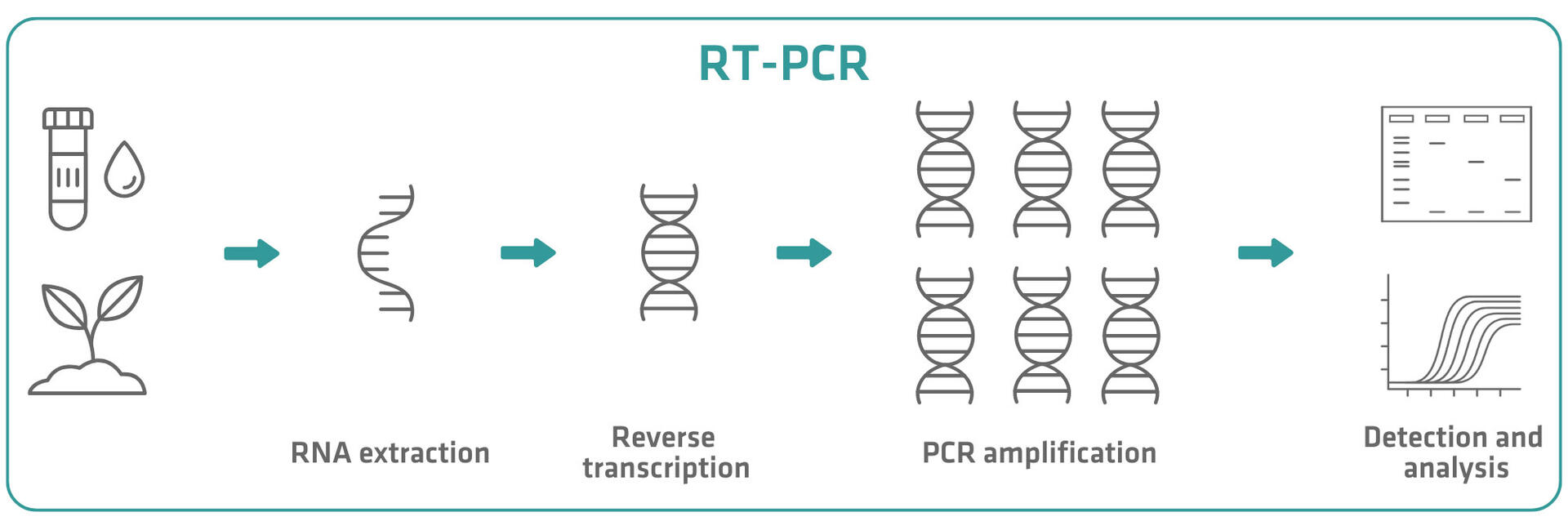
RT-PCR can be divided into 4 steps.
- RNA extraction: For RT-PCR, you first need to extract RNA from biological samples. This is achieved through cell lysis – which releases RNA from within the cells – and the inactivation of RNases to prevent degradation of the RNA molecules. The RNA is then separated from other cellular components and purified using magnetic beads, spin columns or phenol-chloroform reagents like TRIzol™.
- Reverse transcription: Next, RNA is converted into complementary DNA (cDNA) using a reverse transcriptase. These enzymes are naturally found in retroviruses such as HIV, where they convert the RNA genome into DNA to integrate into the host genome for replication. In RT-PCR, reverse transcriptases – such as M-MLV, AMV and HIV-1 – are used to synthesize cDNA from the RNA template.
- PCR amplification: The cDNA is amplified through a standard PCR reaction, which involves repeated cycles of denaturation, annealing and extension. For a detailed explanation of PCR, visit: How does PCR work and what are its 3 steps?
Note that you can also perform a qPCR reaction after reverse transcription if you need to quantify the RNA in your samples. This technique, called RT-qPCR, measures cDNA amplification in real time, allowing you to determine the original RNA quantity in the sample (absolute quantification) or to compare levels or changes in RNA concentration between different samples (relative quantification). For a detailed explanation of qPCR, visit: How qPCR works: SYBR® Green vs. TaqMan® - Detection and analysis: The final step involves detecting and analyzing the amplified cDNA, typically using gel electrophoresis to visualize the presence or absence of specific amplicons. However, if you choose to perform a qPCR reaction in step 3, fluorescent dyes or sequence-specific probes will enable real-time monitoring of amplification, and gel electrophoresis is not needed. The differences between RT-PCR and RT-qPCR are explained in more detail in the next section.
Qualitative RT-PCR vs. quantitative RT-qPCR
While RT-PCR and RT-qPCR share the same initial steps of RNA extraction and reverse transcription, they differ in their subsequent processes and the type of information they provide.
RT-PCR is a qualitative or semi-quantitative method. After reverse transcription, traditional PCR amplifies the cDNA, and the amplified products are typically detected at the end of the reaction using gel electrophoresis. This approach is useful for confirming the presence or absence of a specific RNA sequence, and can provide rough estimates of relative abundance after amplification by comparing the intensity of bands among samples or against controls of a known concentration. However, it does not offer precise quantification, and cannot be used to determine the amount of RNA present before amplification.
In contrast, RT-qPCR quantifies RNA levels with high sensitivity, specificity and precision. Following reverse transcription, qPCR monitors cDNA amplification in real time through fluorescent reporters that emit signals proportional to the amount of amplified cDNA. This allows the detection of specific RNA sequences and the calculation of initial RNA sample concentrations. Several chemistries can be employed to quantify cDNA amplification in RT-qPCR. The 4 primary approaches are SYBR Green, TaqMan probes, molecular beacons and Scorpion probes, each with distinct characteristics and applications.
SYBR Green
SYBR Green is a fluorescent dye that intercalates into double-stranded cDNA during the extension phase of the qPCR reaction. Once intercalated, it shows a strong increase in fluorescence. You can accurately quantify the amount of double-stranded cDNA present by measuring this enhanced signal at the end of each thermal cycle.

SYBR Green is more affordable than the 3 alternative chemistries described below. However, as the dye binds to any double-stranded DNA sequence, the method also detects fluorescence emitted from non-specific qPCR products – such as primer dimers and off-target amplicons – which can lead to an overestimation of the target concentration.
TaqMan probes
TaqMan probes are target specific, and can only bind to the cDNA sequence of interest. They feature a fluorescent reporter dye at the 5’ end and a quencher at the 3’ end. In their intact form, the quencher suppresses the reporter’s fluorescence.
During qPCR, the probe hybridizes to the target sequence. When the Taq polymerase enzyme encounters the TaqMan probe during the extension phase, it displaces and cleaves the 5' reporter dye, separating it from the quencher. This separation increases fluorescence in proportion to the amount of qPCR product.

Molecular beacons
Molecular beacons are target-specific, hairpin-shaped probes designed to bind exclusively to the cDNA sequence of interest. They consist of a loop region complementary to the target, flanked by short stem sequences that bring a fluorescent reporter dye and a quencher into close proximity. In their hairpin conformation, the quencher suppresses the reporter’s fluorescence. During qPCR, when the beacon hybridizes to its target sequence, the hairpin structure opens, separating the reporter from the quencher and generating a fluorescent signal proportional to the amount of specific qPCR product present. Molecular beacons, unlike TaqMan probes, remain intact during amplification and can rebind to their complementary sequence in the next thermal cycle.
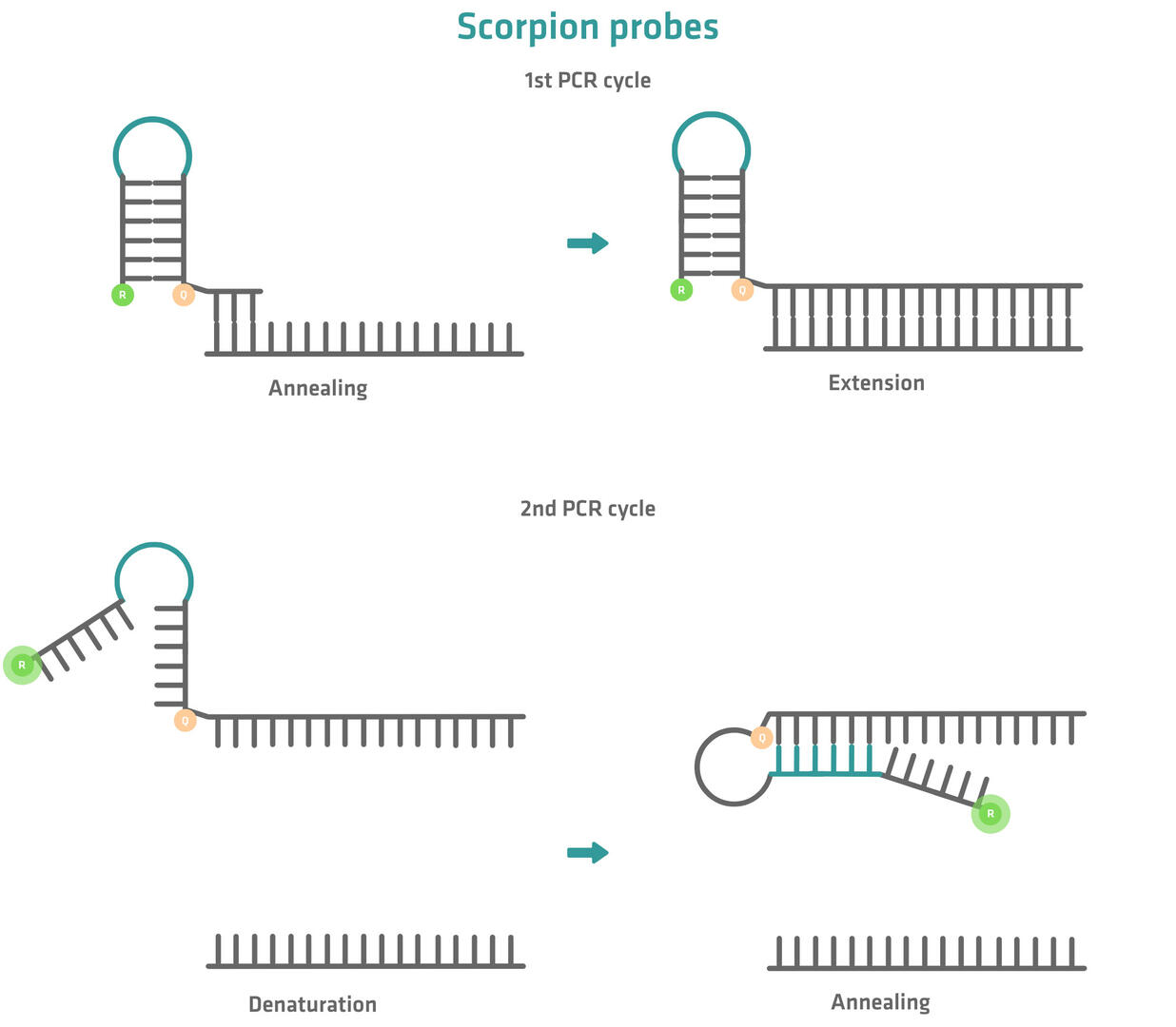
Scorpion probes
Scorpion probes combine primer and probe functions in a single molecule. They comprise a PCR primer linked to a probe sequence that forms a hairpin loop structure. As with molecular beacons, the probe consists of a target-specific sequence and a short stem sequence, ensuring that the fluorescent signal of the reporter dye is suppressed by a quencher.
During the first PCR cycle, the primer portion of the Scorpion probe anneals to the target sequence. The Taq polymerase enzyme then synthesizes a new, complementary strand.
During the second PCR cycle, the hairpin structure opens and the sequence-specific part of the Scorpion probe binds to the previously synthesized strand. During this process, the reporter is separated from the quencher and a fluorescent signal is generated.
Choosing the right detection chemistry depends on your specific application. SYBR Green is the most affordable option, whereas TaqMan probes, molecular beacons and Scorpion probes offer high specificity. Moreover, they can be used to perform multiplex assays, as you can label different probes or beacons with different fluorescent dyes.
1-step vs. 2-step RT-PCR
Besides the distinction between qualitative RT-PCR and quantitative RT-qPCR, there is another key differentiation between 1-step and 2-step RT-PCR. The following sections outline how these 2 reaction types work, along with their advantages and disadvantages. The same principles apply to 1-step and 2-step RT-qPCR, the only difference being the addition of a fluorescent reporter to the reaction mixture.
2-step RT-PCR
Reverse transcription and PCR are performed separately in 2-step RT-PCR. RNA is first converted into cDNA using reverse transcriptase enzymes and oligo-dTs, random oligomers or gene-specific primers. The cDNA sequences are then amplified by polymerase enzymes along with gene-specific primers.

This approach offers flexibility in primer selection, as oligo-dTs, random oligomers, gene-specific primers or a combination of these can be used for reverse transcription.
- Oligo-dTs selectively reverse transcribe mRNA into cDNA, making them the preferred option for creating complete mRNA libraries for downstream applications, such as transcriptional analysis.
- Random oligomers generate a cDNA library from the entire RNA population, but they produce shorter cDNA fragments than the other options, as synthesis begins at multiple points within an RNA molecule.
- Gene-specific primers enable the targeted reverse transcription of specific genes.
The ability to use oligo-dTs and random oligomers makes it possible to generate a cDNA library containing a broad range of sequences. This library can then be used in multiple, independent PCR reactions to analyze different target genes from the same sample.
Beyond primer flexibility, 2-step RT-PCR offers additional advantages. Since reverse transcription and PCR are conducted separately, each step can be optimized independently, improving reaction sensitivity and efficiency – an important factor when working with limited RNA samples. Furthermore, the cDNA library created by reverse transcription can be stored for future use, allowing researchers to validate results or analyze additional targets from the same RNA sample later on.
The primary drawbacks of 2-step RT-PCR are its increased time requirements and the need for more pipetting steps. These factors make the method less automation friendly, while also raising the risk of contamination and result variability.
1-step RT-PCR
In a 1-step RT-PCR reaction, reverse transcription and amplification occur within a single tube. First, gene-specific primers and reverse transcriptase enzymes convert RNA into cDNA. The same gene-specific primers, along with polymerase enzymes, then amplify the cDNA sequences.
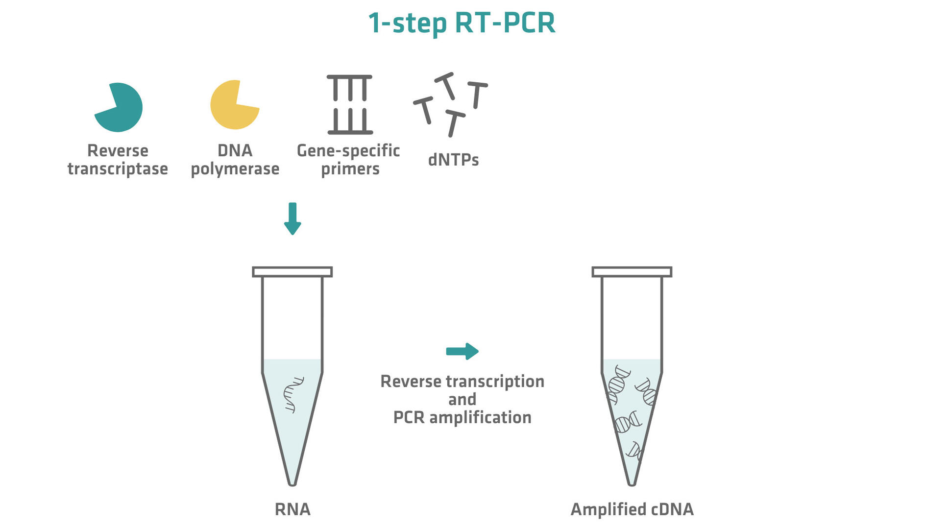
Consolidating both reactions makes 1-step RT-PCR faster, with less hands-on time. It also minimizes the contamination risk, as the sample remains untouched after reverse transcription, while the reduced number of pipetting steps further decreases the chance of errors and variability. This method is ideal for high throughput applications, as the simplified protocol makes automation easier.
However, 1-step RT-PCR is best suited for experiments targeting only a few genes. Since gene-specific primers are required for the reverse transcription step, it is not possible to generate a comprehensive cDNA library representing all mRNAs or total RNA. Moreover, because reverse transcription and PCR occur in the same tube, there is no opportunity to optimize the reaction conditions separately, which may lead to lower reaction efficiency and yields. Another limitation is that the cDNA produced in the reverse transcription step is immediately consumed in the PCR process, preventing its storage for future validation or for targeting additional genes from the same sample.
In summary, 1-step RT-PCR is best suited for high throughput applications with many samples but only a few target genes. In contrast, 2-step RT-PCR is the preferred choice when analyzing multiple targets from the same RNA sample, which requires high sensitivity and efficiency, or long-term cDNA storage for future experiments.
How to avoid DNA contamination
DNA contamination in RT-PCR experiments can lead to false positive results. For instance, consider a scenario where you aim to determine whether a specific gene has been transcribed into mRNA in your sample. If the gene was not transcribed, no mRNA for that gene should be present. However, if gDNA contamination occurred during RNA extraction, gDNA corresponding to that gene might be present. During the RT-PCR process, this contaminating gDNA could be amplified, leading to a false positive result that incorrectly suggests the gene was transcribed.
To minimize the risk of gDNA contamination and ensure reliable results, 2 strategies can be employed: treating the sample with a DNase, and designing primers that span an exon-exon junction or flank an intron.
DNase treatment
The first option is to enzymatically remove gDNA contamination by incorporating a DNase digestion step after RNA extraction and before reverse transcription. Adding a DNase enzyme (e.g. DNase I) to the purified RNA sample selectively degrades single- and double-stranded DNA without harming RNA. The DNase can then be inactivated using a heat treatment or DNase removal agent. Digestion efficiency can be validated using no-reverse transcription (no-RT) controls. No-RT controls are negative controls that skip the reverse transcription step before PCR amplification. If amplification occurs in the no-RT control, it indicates that there is still contaminating gDNA in the sample.
RT-PCR primer design
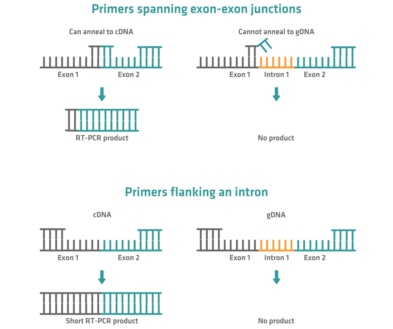
If you want to analyze mRNAs from eukaryotic organisms, there is a second option to ensure that gDNA has no impact on the subsequent PCR or qPCR. Many eukaryotic genes contain introns (non-coding DNA sequences). When such a gene is transcribed into mRNA, these introns are removed from the final mature mRNA in a process called splicing.
This means that primers spanning an exon-exon junction will be able to anneal to cDNA from mRNA molecules, but not to gDNA molecules. Alternatively, primers can be designed to span large introns. Although they can anneal to cDNA and gDNA, only cDNA will be amplified during PCR, resulting in short amplicons. gDNA won't be amplified because the length of the amplification step can be set to be too short to finish amplification of the much longer gDNA sequences.
Applications
RT-PCR and RT-qPCR have become indispensable tools in gene expression studies, genetic engineering and clinical diagnostics. Moreover, they are used in forensic science, food safety and agriculture, environmental research and monitoring, and drug discovery. This section lists some of the most common applications.
Gene expression studies
RT-PCR and RT-qPCR are considered the gold standard for gene expression analysis due to their high sensitivity and specificity. They allow researchers to detect and quantify mRNA levels to understand how genes are switched on or off in different tissues, developmental stages or disease conditions.
Genetic engineering
RT-PCR is a useful tool for genetic engineering applications, enabling researchers to clone specific genes from mRNA. These cloned genes can then be used to create genetically modified cell lines or organisms. For example, the genome of AquAdvantage salmon contains a growth hormone gene isolated from Chinook salmon pituitary gland mRNA, promoting rapid growth during early life.1
Clinical diagnostics
RT-PCR and RT-qPCR are also used in clinical diagnostics to detect and monitor infectious diseases, cancer biomarkers and genetic disorders.
- Viral detection: Both methods are widely used to identify RNA viruses such as SARS-CoV-2, HIV and hepatitis C. Their high sensitivity and specificity allow early infection detection. This was particularly crucial during the COVID-19 pandemic, when the rapid identification of infected individuals – ideally before they exhibited symptoms – played a key role in controlling the spread of disease through timely isolation measures.
- Cancer diagnostics: Many cancers exhibit distinct RNA-based biomarkers. RT-PCR and RT-qPCR enable the detection and quantification of these markers, aiding in early diagnosis and personalized treatment strategies.
- Genetic disorder screening: By analyzing gene expression patterns, RT-PCR and RT-qPCR help in diagnosing hereditary diseases and monitoring gene mutations associated with genetic disorders. For example, they are widely used in screening for cystic fibrosis.
Forensic science
RT-PCR and RT-qPCR have proven valuable in forensic investigations by enabling the analysis of RNA in crime scene samples. For example, experts can estimate the age of bloodstains and other biological materials by examining degradation patterns, providing crucial timeline information. Furthermore, the ability to detect specific RNA markers has improved the accuracy of body fluid identification, enabling forensic teams to differentiate between blood, saliva, semen and other bodily fluids.
Food safety and agriculture
RT-PCR and RT-qPCR are vital tools for monitoring pathogens, ensuring food safety and protecting crop health in food and agricultural sciences. For instance, they enable the rapid and precise detection of foodborne bacteria like Salmonella, Listeria and Escherichia coli, helping to prevent outbreaks. Additionally, RT-PCR is widely used to identify and quantify genetically modified organisms in food and feed products, ensuring compliance with regulatory standards. In agriculture, it aids in the early detection of viral and bacterial infections in crops, allowing timely intervention.
Environmental research and monitoring
RT-PCR and RT-qPCR are playing a growing role in environmental science and ecology, enabling the detection and analysis of microorganisms in various ecosystems. They help researchers to study microbial communities in water, soil and air, providing insights into their composition and dynamics to support biodiversity studies and conservation efforts. Moreover, these techniques are crucial for identifying pathogens and indicator organisms to assess water quality in drinking water, recreational areas and wastewater treatment systems.
Drug discovery
RT-PCR and RT-qPCR play crucial roles in various stages of drug development.
- Target validation: They help researchers to confirm the involvement of specific genes or proteins in disease processes, validating them as potential drug targets.
- Efficacy testing: They are used to assess how candidate drugs affect gene expression, providing insights into their mechanisms of action and effectiveness.
- Toxicity studies: They aid in evaluating the potential side effects of drugs by analyzing changes in gene expression patterns in response to drug exposure.
Conclusion
RT-PCR has firmly established itself as an indispensable technique in molecular biology and diagnostics, allowing researchers and clinicians to detect even minimal amounts of RNA with high accuracy. Whether used in qualitative or quantitative formats, as 1-step or 2-step approaches, each method offers specific advantages depending on the question to be answered.
Are you using RT-PCR in your work, or do you have questions about its applications? Share your thoughts in the comments!
Ask our expert. Leave a comment!
Write us if you have any questions regarding the blog article.