-
Automation-enabled high throughput electroporation of primary cells using the ASSIST PLUS and DropGenie platform
Precision workflow for low-cell-input transfection
There is increasing demand for scalable and reproducible gene editing of clinically-relevant cell types for the development of new therapies. Combined with the limited availability of patient samples, this is driving a need for automated platforms capable of minimizing the amount of cellular material required. However, traditional electroporation approaches are difficult to miniaturize and poorly suited for high throughput automation.
The DropGenie Transfection Platform combines digital microfluidics (DMF) and tri-drop electroporation to enable efficient genome editing with drastically reduced cellular input requirements. This revolutionary approach decreases the number of cells needed in one experiment from millions to thousands per edit, greatly benefiting CRISPR screening in rare or precious cell types.
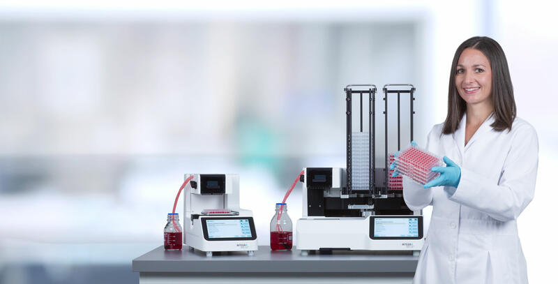
INTEGRA's ASSIST PLUS pipetting robot and VOYAGER adjustable tip spacing pipette enable automated liquid handling for DropGenie workflows. Together, DropGenie’s microfluidic electroporation technology and INTEGRA’s automation provide a gentle, high throughput solution for genome editing of precious primary cells.
-
Table of contents
There is increasing demand for scalable and reproducible gene editing of clinically-relevant cell types for the development of new therapies. Combined with the limited availability of patient samples, this is driving a need for automated platforms capable of minimizing the amount of cellular material required. However, traditional electroporation approaches are difficult to miniaturize and poorly suited for high throughput automation.
The DropGenie Transfection Platform combines digital microfluidics (DMF) and tri-drop electroporation to enable efficient genome editing with drastically reduced cellular input requirements. This revolutionary approach decreases the number of cells needed in one experiment from millions to thousands per edit, greatly benefiting CRISPR screening in rare or precious cell types.
INTEGRA's ASSIST PLUS pipetting robot and VOYAGER adjustable tip spacing pipette enable automated liquid handling for DropGenie workflows. Together, DropGenie’s microfluidic electroporation technology and INTEGRA’s automation provide a gentle, high throughput solution for genome editing of precious primary cells.
Key benefits
- Seamless integration with assay formats: The combination of VIALAB programming and ASSIST PLUS enables gentle, automation-ready handling of sensitive primary cells.
- Gentle handling of precious samples: The ASSIST PLUS and VOYAGER pipette provides full control over pipetting speed and height, enabling bubble-free transfers that preserve cell integrity.
- High delivery and knockout performance with small volumes: The DropGenie platform miniaturizes electroporation, enabling robust GFP delivery, as well as efficient CRISPR knockouts with excellent recovery and viability.
- User-friendly and high-throughput: The VOYAGER pipette’s electronic adjustable tip spacing, reduces setup time, enabling hundreds of edits per hour.
- Accessible and easy to use: Combining a compact footprint that fits directly into a tissue culture hood and a simple to operate workflow, makes high-quality genome editing accessible to any lab.
Overview: How to load the DropGenie with the ASSIST PLUS
In this application note, we demonstrate the automated loading and offloading of precious adherent or suspended primary cells on the DropGenie electroporation platform using the ASSIST PLUS pipetting robot. The 8 channel, 125 μl VOYAGER adjustable tip spacing pipette automates all liquid handling steps for loading and recovery, while the DropGenie dock and cartridge enable gentle cell handling for consistent generation of cell packets, rapid ribonucleoprotein (RNP) formation and gentle electroporation. Figure 1 illustrates the step-by-step procedure of the automated workflow for arrayed CRISPR screening protocols.
Downloads: App note and protocols for automation-enabled high throughput electroporation of primary cells using the ASSIST PLUS and DropGenie platform
Experimental set-up
Step-by-step procedure
Cell culture and preparation
- For primary fibroblast-like synoviocytes (FLS), a standard operating procedure for adherent cell lines was followed as described in Patel et al. (2025).1
- Primary human T-cells were prepared according to the protocol described by Patel et al. (2025).1
- Jurkat cell suspensions were cultured and harvested following the protocol published by Little et al. (2023).2
Electroporation workflows
1. Cell counting experiments
- Input: 45 µl of prepared cells were loaded into wells C1 and F1 of the source plate.
- Buffer handling: columns 2-10 were filled with fluorescence-activated cell sorting (FACS) buffer for acute cell counting via flow cytometry, enabling direct offloading into the FACS buffer and minimizing cell losses due to washing.
2. GFP mRNA delivery
- Input: 45 µl of prepared cells were loaded into wells C1 and F1 of the source plate.
- Plate type: round-bottom plates were used for cell suspensions; flat-bottom plates were used for adherent cells.
- Reagent: cells were spiked with the desired concentration of GFP mRNA.
- Collection wells: columns 2-10 were filled with complete medium.
3. CRISPR knockout experiments
- Input: 45 µl of prepared cells were loaded into wells C1 and F1 of the source plate.
- Plate type: round-bottom plates were used for cell suspensions; flat-bottom plates were used for adherent cells.
- Reagents: cells were spiked with the appropriate concentration of Cas9 protein.
- Single guide RNA (sgRNA) delivery: target sgRNAs were pre-deposited onto the cartridge bottom plate using a nanodispenser prior to cartridge assembly.
- Collection wells: columns 2-10 were filled with complete medium.
Cartridge assembly and run execution
- Cartridge assembly may occur at any time prior to use for experiments without reagent deposition.
- Cartridge assembly should be performed immediately after nanodispensing for experiments with reagent deposition.
- The operator should wear gloves to snap together the cartridge bottom and top plates, ensuring correct orientation.
- Initiate the DropGenie program by manually inputting the electroporation parameters, or by importing a .CSV file with the electroporation parameters.
- Then position the assembled cartridge on the DropGenie dock.
Pre-electroporation: loading the cartridge with the ASSIST PLUS
Note: Tips in rows A, H, I and P of the tip rack MUST be removed before the run (Figure 3).
Although the VIALAB program displays 8 positions per column on the DropGenie cartridge, only 6 exist. The program is adjusted for the 8 channel pipette to perform the transfers correctly. Rows A, I H and P (marked with an X in subsequent figures) are left empty.
Transfer the cells and liquid electrode onto the DropGenie cartridge using the 8 channel, 125 µl VOYAGER pipette mounted on the ASSIST PLUS, select the protocol and allow the ASSIST PLUS to idle. Prior to running the protocol, ensure that the DropGenie program has been initiated, and is prompting ‘Load Reagents’. At the same time, the light on the front of the DropGenie system should be flashing. Run the VIALAB program 'Drop-Genie_plate_fill'. After 3 mixing cycles to ensure a uniform suspension of cells, 9.2 µl of liquid electrode (wells B1, D1, E1 and G1) and 9.2 µl of cells with additional payload (wells C1 and F1) are transferred from the 96 well round-bottom microplate at deck position B into columns C1, C4, C9 and C12 of the DropGenie cartridge at deck position C. To guarantee pipetting precision, a 5 µl pre-dispense and a 4 µl post-dispense are applied (Figure 4).
Electroporation: running the DropGenie System
Once all the reagents have been fed into the cartridge, proceed to the next step on the DropGenie software by pressing ‘Enter’. The DropGenie liquid handling should start and run these steps:
a. Droplets containing cell suspensions (~48 units, each with fewer than 10,000 cells mixed with Cas9) are dispensed onto the array.
b. Guide RNAs deposited on the substrate are rehydrated through pulsed droplet mixing for ~1 minute.
c. On-chip RNP complexes form during a brief incubation period.
d. Electroporation is carried out at each individual site, with user-adjustable parameters.
e. Tri-drops are transported to the designated offloading ports for collection.
Post-electroporation: offloading the cartridge
After the DropGenie performs electroporation, 14.5 µl of cell medium in wells B2-G2 is transferred from the 96 well round-bottom microplate at deck position B, into columns C2, C3, C5, C6, C7, C8, C10 and C11 of the DropGenie cartridge at deck position C to quench the samples. A 5 µl pre-dispense and a 2 µl post-dispense are applied (Figure 5).
Finally, the transfected cells are mixed 3 times with the cell medium to ensure no cells remain in the tip, and 22.5 µl is transferred into columns 3-10 of the 96 well round-bottom microplate at deck position B (Figure 6). Once offloading is completed by the ASSIST PLUS, the protocol on the DropGenie system can be completed by clicking through the prompts. Cells are then placed in an incubator, and phenotypic or genomic analysis can be performed as required.
Results
This application note evaluated the compatibility of the ASSIST PLUS with the DropGenie system by measuring cell recovery for both cell suspensions and adherent cells. Cells were washed and resuspended in DMFection buffer prior to being loaded into the cartridge. Cells were either dispensed directly into the offloading plate as a 1 μl off-chip control or loaded into the DropGenie cartridge by the ASSIST PLUS and run through the electroporation workflow at either 0 V or 500 V before automated offloading into a 96 well plate. Acute cell counts were then measured immediately after offloading using flow cytometry for Jurkat and primary T-cells, or microscopy for FLS.
For Jurkat cells resuspended at 10 million/ml, counts were consistent across all conditions: manual off-chip controls, automated loading/offloading with no electroporation, and automated loading/offloading with 500 V electroporation. The CV in the 500 V condition was 12.7 % (n=37), demonstrating highly reproducible recovery even at low cell numbers. Similar results were observed for FLS, where microscopy-based counts showed no significant differences across conditions. Here, the CV was higher at 35.0 % (n=38), as expected for microscopy-based methods that capture only a portion of the well. To assess reproducibility across cartridges, we compared primary T-cell recovery using 3 independent cartridges loaded from the same source population and observed no significant differences. The overall CV across cartridges was 16.7 % (n=115).
Together, this data demonstrates that the ASSIST PLUS and DropGenie workflow yields robust and consistent cell recovery across cell suspensions and adherent cell types. It does not introduce additional variability, or result in a loss of cells during automated handling on the ASSIST PLUS or DropGenie platform. However, these measurements were acute and viability was not assessed separately.
After demonstrating the robustness of the workflow across multiple cell types and conditions, we next evaluated GFP mRNA delivery in Jurkat cells. Cells were mixed with GFP mRNA, loaded into the DropGenie cartridge, and subjected to 500 V electroporation before automated offloading. After 24 hours of incubation, flow cytometry analysis showed highly efficient delivery, with mean transfection >95 %, CV <5 %, and viability >95 % across 39 independent electroporation experiments. These results confirm the effectiveness of the platform for delivering nucleic acid cargoes into cells using electroporation.
We next assessed GFP mRNA delivery into primary adherent FLS. Electroporation was performed at 500 V with inputs of ~500, 1,000 or 5,000 cells per experiment to highlight the platform’s ability to handle low cell numbers. Confluency profiles were comparable between electroporated and non-electroporated wells for 6 days, with all conditions showing peak GFP expression at ~24 hours after electroporation. Delivery efficiency reached ~95 % in the 1,000 and 5,000 cell conditions, while the 500 cell condition peaked closer to 70 %, likely reflecting reduced uptake efficiency at sparse densities or limited availability of GFP per tri-drop. Standard deviation also increased at lower inputs, consistent with greater variability in smaller populations. Microscopy confirmed robust GFP expression across all conditions, with morphology suggesting healthier cells at higher densities.
Table 1: Descriptive statistics of GFP confluence over time for FLS after 24 hours.
We also evaluated knockout of the TRAC locus in primary T-cells to demonstrate the ability of this workflow for arrayed CRISPR screening. TRAC knockout was chosen as a model because TCR α/β is a surface marker readily quantifiable by flow cytometry, enabling rapid and reproducible assessment of editing efficiency.
sgRNAs targeting TRAC or scramble controls were deposited across the cartridge using an automated nanodispenser, and cells were processed through the full ASSIST PLUS-DropGenie workflow. The workflow yielded robust gene disruption with high repeatability across multiple independent experiments. Acute viability remained >80 % in all conditions, confirming that automated loading, electroporation and offloading are well tolerated. TRAC knockout efficiency was 92.5 % in CD4⁺ T-cells and 90.9 % in CD8⁺ T-cells, with CVs below 5 %, underscoring the precision and reproducibility of the integrated workflow, even when operating at low cell inputs. These findings establish the combined ASSIST PLUS-DropGenie set-up as a powerful solution for arrayed CRISPR screening in primary human T-cells, with the potential to scale up towards larger gene panels and diverse donor-derived samples.
References
- Patel, M.A., Boribong, B.P., Sinha, H. et al. Miniaturized scalable arrayed CRISPR screening in primary cells enables discovery at the single donor resolution. Sci Rep 15, 29350 (2025). https://doi.org/10.1038/s41598-025-13532-z
- Little, S.R., Leung, Z., Quach, A.B.V., Hirukawa, A., Gholizadeh, F., Hajiaghayi, M., Darlington, P.J. and Shih, S.C.C. (2023), A Tri-Droplet Liquid Structure for Highly Efficient Intracellular Delivery in Primary Mammalian Cells Using Digital Microfluidics. Adv. Mater. Technol., 8: 2300719. https://doi.org/10.1002/admt.202300719
Conclusion
- Consistent recovery across cell types: the ASSIST PLUS and DropGenie workflow maintained reproducible cell counts in both suspension and adherent formats, confirming delicate automated loading of fragile cells.
- Robust GFP delivery and reproducibility: electroporation of GFP mRNA achieved highly efficient and repeatable delivery in this automated setting.
- Low input compatibility: reliable GFP expression was achieved even at inputs as low as 500 cells per experiment, enabling work with scarce or precious samples.
- Scalable knockout performance: arrayed CRISPR experiments produced high knockout efficiencies with excellent viability and low variability, validating the combination of ASSIST PLUS and DropGenie for functional genomics applications.
Ask our expert. Leave a comment!
Write us if you have any questions regarding the application note or one of our instruments.
Any questions? I'm happy to help!
Instruments and accessories
ASSIST PLUS, Pipetting Robot
INTEGRA has developed the ASSIST PLUS pipetting robot to streamline routine pipetting tasks at an affordable price. Using INTEGRA electronic multichannel pipettes, the system:
- automates pipetting tasks,
- eliminates physical strain and
- ensures superior reproducibility and
- error free pipetting.
Part No. 4505
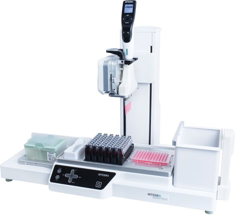
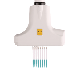
VOYAGER, 8 Channel, 125 µl, electronic pipette
VOYAGER pipettes allow the tip spacing to expand anywhere between 4.5 mm and 33 mm at the push of a button.
- Single handed operation leaves the other hand free to handle labware.
- On the fly access to up to 3 user-defined tip spacings.
- These user defined tip spacings are saved. No need to memorize spacings of different labware formats.
Part No. 4722
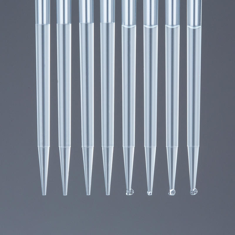
GRIPTIPS®, 125 µl, Sterile, Filter, Low retention
INTEGRA uses a unique polypropylene blend to mold Low Retention GRIPTIPS® with heightened hydrophobic properties. Using Low Retention GRIPTIPS® prevents low surface tension samples from spreading out and “wetting” the inner wall of the tips, allowing them to bead-up for a maximum liquid recovery.
Part No. 6565
DropGenie: DropGenie platform
Miniaturize and automate workflows at scale—to extract deep insights from every drop.
The platform redefines lab automation—enabling high-throughput, low-volume workflows that turn precious patient samples into rich, high-dimensional datasets—accelerating genomic discovery and powering the future of personalized medicine.
Source: Supplier Website
DropGenie: DropGenie cartridge
Miniaturize and automate workflows at scale—to extract deep insights from every drop.
The platform redefines lab automation—enabling high-throughput, low-volume workflows that turn precious patient samples into rich, high-dimensional datasets—accelerating genomic discovery and powering the future of personalized medicine.
Source: Supplier Website
DropGenie: DMFection buffer
Miniaturize and automate workflows at scale—to extract deep insights from every drop.
The platform redefines lab automation—enabling high-throughput, low-volume workflows that turn precious patient samples into rich, high-dimensional datasets—accelerating genomic discovery and powering the future of personalized medicine.
Source: Supplier Website
DropGenie: 1% Surfactant F
Miniaturize and automate workflows at scale—to extract deep insights from every drop.
The platform redefines lab automation—enabling high-throughput, low-volume workflows that turn precious patient samples into rich, high-dimensional datasets—accelerating genomic discovery and powering the future of personalized medicine.
Source: Supplier Website
Corning: Falcon 96 well clear round-bottom microplate
- Round bottom wells with 330 µL total volume
- Recommended working volumes of 75 to 200 µL
- Treated for optimal cell attachment
- Sterilized by gamma radiation and nonpyrogenic
- Individual alphanumeric codes for well identification
- Bulk packed 20 per bag
Source: Supplier Website
Downloads
Download App Note as PDF
DownloadAutomation-enabled high throughput electroporation of primary cells using the ASSIST PLUS and DropGenie platformCustomer’s voice
We’ve been using the Assist Plus for over two years, and it has been extremely valuable for setting up high-throughput assays. With the 4-deck configuration, plates must be removed manually when running larger batches, but the system is excellent for mid-throughput workflows. It’s very straightforward to set up and operate. One consideration is that only specific tips are compatible with the instrument, which adds an additional cost to factor in. Overall, we’ve been very happy with the setup!