
Tips for preventing contamination in cell culture
The discovery of contaminated samples is costly for any scientist, as valuable reagents and research time are lost and, in worse case scenarios, entire batches can be spoiled. However, if undetected, contamination may cause unexpected or inaccurate conclusions to be drawn about the behavior and survival of cells. This can pose a potential health and safety risk if contamination is not detected in the research and development of vaccines and biopharmaceuticals. All apparent cell culture conditions need to be reproducible for cell-based therapies to be used as effective tools, but this is not possible with the hidden influence of contamination.
The hidden risk of cell contamination
The consequences of contamination can range from minor inconveniences, such as the need to dispose of a contaminated flask of cells, to wider consequences, such as the publication of supposedly trusted results that are inaccurate due to undetected cross-contamination. It is estimated that more than 15 % of cell culture studies are based on misidentified or cross-contaminated cell lines that can go entirely undetected, compromising the reproducibility and accuracy of results.1 For example, some incidences may influence the behavior of cells – including metabolism and growth – that, if undetected, may lead you to mistakenly believe that you have identified a new cell line or uncovered a new innate behavior. Therefore, a lot of our cell culture predictions and beliefs may be based on false information, from cell lines without the correct authentication.
Biological contamination
Staggeringly, the incidences of biological contamination are reportedly far more common, as most recurring problems are expected to be microbial in origin.2 Unfortunately, the conditions needed to culture healthy cells – such as the nutrient-rich media and controlled temperatures – are also the perfect environment for the growth and proliferation of some unwanted microorganisms. Biological contamination of samples can be described as the in vitro equivalent of an emerging disease; sometimes cells must compete for essential nutrients and defend themselves against toxic byproducts as invading microbes grow and replicate,3 while some contaminants can engulf entire cells. In addition, mycoplasma contaminants have been proven to modify gene expression to disrupt important cellular pathways.4 This has a huge impact on the viability of cells, resulting in poor cell health and death. How visible an infection is to the naked eye, or even under the microscope, often determines how likely it is to go undetected. Here are a few common contaminants to avoid, and how to identify them:
- Bacteria – A bacterial infection usually leaves cells looking particularly unhealthy, causing a swift change in turbidity or color of the media. Moving bacteria may also be visible through a microscope.
- Fungus – Fungal contaminants, such as molds and yeasts, often grow into clearly visible structures in media, which can be readily detected by the naked eye or under a microscope.
- Mycoplasma – Mycoplasma are the smallest of all free-living organisms, and are not usually visible, even under a microscope. They also lack a cell wall and are, therefore, immune to the activity of antibiotics. Specific testing, such as with PCR or fluorescent DNA staining, is the only way to detect any contamination.
Aseptic working techniques and sterile environments can help to reduce the risk of the biological contamination of cells. Spoiled cell samples or reagents need to be correctly disposed of and, even in minor incidences of contamination – such as opening a single flask containing airborne microorganisms under laminar flow – requires the entire environment to be decontaminated to prevent particles from settling. Specific testing for contaminants, along with regular microscopic observations, should be adopted as a standard quality control screening procedure in all cell culture workflows. In addition, the FDA actively encourages biopharmaceutical manufacturers to test the quality of cell substrates.5

Aseptic procedures to prevent contamination
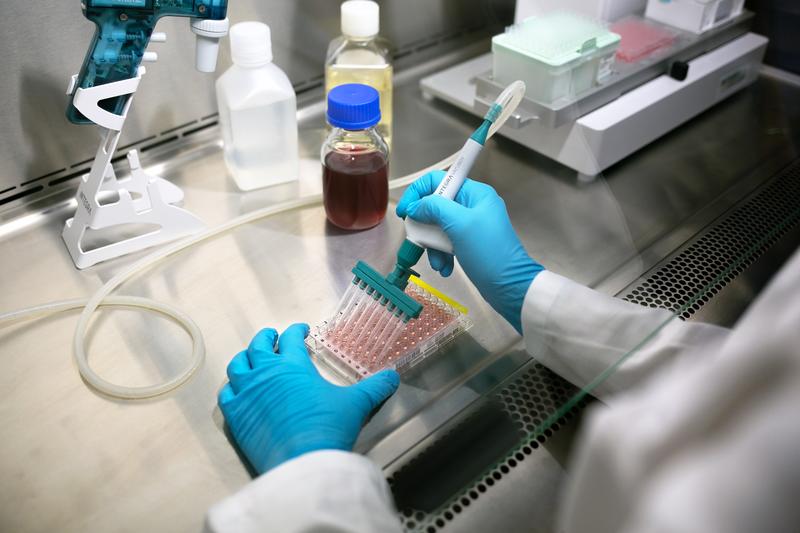
Unfortunately, it is nigh on impossible to completely eliminate all sources of contamination, but there are various aseptic procedures that can be adopted to minimize the occurrence – beginning with using the correct tools. Here are some suggestions for selecting the correct pipettes and tips, and using proper handling for cell culture workflows:
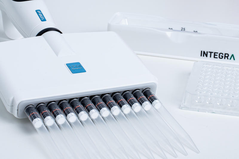
- Sterile pipettes and tips should always be used for cell culture. Tips should come with a statement of sterility when they are purchased, to alleviate concerns of biological contamination.
- Filter tips should be used to prevent aerosols created during aspiration from entering the barrel of the pipette, which could result in the cross-contamination of cell lines in future samples.
- Every tip should form a perfect seal with the pipette, and never leak or fall off. This prevents liquid from spilling or dripping on the workbench, risking contaminating other samples or the lab worker.
- Proper pipetting procedures should include pre-wetting techniques. Aspirating and expelling the full volume of the pipette with the liquid prior to pipetting the sample is generally good pipetting practice, to equilibrate the tip environment and reduce the risk of tip dripping.
- Automation is the best solution to reduce the risk of contamination from the user, as it is hands-off. It also helps to reduce human error and the likelihood of the cross-contamination of cell lines, improving the reproducibility of tasks. Ideally, the automation set-up should be protected by a well-maintained laminar flow cabinet, and subjected to frequent cleaning procedures.
Check out how INTEGRA can help you to improve your cell culture handling.
Further reading: An introduction to cell culture and its challenges
References
- Nardone, R. M. (2008). Curbing rampant cross-contamination and misidentification of cell lines. Biotechniques, 45(3), 221-227.
- Langdon, S. P. (2004). Cell culture contamination. Cancer Cell Culture, 309-317.
- Lincoln, C. K., & Gabridge, M. G. (1998). Cell culture contamination: sources, consequences, prevention, and elimination. Methods in cell biology, 57, 49-65.
- Chernov, V. M., Chernova, O. A., & Sanchez-Vega, J. T. (2014). Mycoplasma contamination of cell cultures: vesicular traffic in bacteria and control over infectious agents. Acta Naturae (англоязычная версия), 6(3 (22)).
- U.S. Department of Health and Human Services. (2018). Food and Drug Administration. Elimination of the 21 CFR 610.30 Test for Mycoplasma, 1-6.