From lysate to pure protein: a practical guide to protein purification methods
Written by Anina Werner · 28. July 2026
In January 1922, a 14-year-old boy dying of diabetes became the first person to be treated with purified insulin. The protein itself had been known for years, but turning a crude pancreatic extract into something pure and safe enough to inject into a patient was the real breakthrough.1
A century later, protein purification is central to research, biotechnology and medicine, whether the goal is to study protein-protein interactions, produce enzymes for food processing or manufacture a therapeutic antibody.
This blog discusses the general protein purification workflow, compares the most widely used techniques and explains how researchers verify that their final sample is pure, concentrated and functional. We'll also cover areas where protein purification is applied, starting at basic research all the way to biopharmaceutical manufacturing.
Table of contents
What is protein purification?
Protein purification is the process of isolating a specific protein of interest from a complex biological mixture – such as a cell culture or tissue sample – by removing contaminants like other proteins, nucleic acids, lipids and small molecules.
Protein purification is sometimes also called protein isolation, although some researchers reserve ‘isolation’ for the initial extraction of the protein from its biological source, and refer to ‘purification’ as the subsequent steps that increase its purity. In practice, this is usually treated as a single, continuous workflow rather than 2 separate operations and, in this article, we will use the term ‘protein purification’ to refer to the process as a whole.
Protein purification challenges
Protein purification comes with several difficulties. One of the most fundamental challenges is the immense diversity of proteins; they differ in size, charge, solubility, hydrophobicity and binding affinity, meaning that no single purification method is suitable for every target. Instead, researchers typically need to combine multiple techniques – each exploiting a different property of their protein of interest – to progressively remove contaminants.
Another major challenge when setting up a protein purification workflow is balancing purity and yield. Each additional clean-up step helps to achieve higher purity, but results in a lower recovery rate of the target protein. This can be problematic because proteins are often present in limited quantities and cannot be amplified. The starting amount represents the maximum material available, and researchers must carefully determine how much purity is actually required for the intended downstream application.
A key question that should also be addressed at the very beginning is whether the target protein needs to remain in its native, correctly folded structure so that it retains its biological function. If so, purification must be carried out under gentle, non-denaturing conditions that preserve stability, potentially limiting the range of methods that can be used.

The number of purification steps and methods chosen may vary widely based on the target protein, as well as the purity and functionality requirements of downstream applications. However, most workflows follow a broadly similar sequence of stages.
- Sample collection/production: the target protein is either extracted directly from its natural source (tissue, body fluid or unmodified cell culture sample) or produced recombinantly by a host expression system, such as E. coli, yeast, insect or mammalian cells. In the recombinant case, you first need to engineer cells with a vector encoding the protein of interest, and then culture them under conditions that induce expression. Recombinant expression enables high yields and the use of tags that simplify downstream purification. However, native samples are still the preferred choice when it’s important to preserve the protein’s natural context, or when the target protein is difficult to express recombinantly.
- Extraction: cells are disrupted using mechanical, chemical and/or enzymatic methods to release their contents, including the target protein, into a solution. Proteins are inherently unstable, vulnerable to aggregation, degradation by proteases, and denaturation from temperature or pH changes. Buffers used for this step therefore typically contain protease inhibitors, and extraction is performed at a low temperature, while maintaining suitable pH and ionic strength.
- Purification: the target protein is isolated from other cellular components using one or more purification techniques.
- Characterization and quantification: the purified protein is analyzed to confirm its identity and determine its purity, concentration and, if necessary, biological activity before use or storage.
The following sections explore the different techniques for steps 3 and 4 in more detail.
Protein purification techniques
Researchers typically combine several purification methods to isolate a specific protein. The optimal workflow depends on the protein’s properties, the required purity, and the planned downstream applications. Common methods used include centrifugation, precipitation, chromatography, ultrafiltration and dialysis. In this section, we will describe these methods and explain when they are typically used.
Centrifugation
Once cells have been disrupted, the resulting mixture – the lysate – contains contaminants of different size and density alongside the target protein. Centrifugation is therefore often used as an initial, cost-effective purification method. Low-speed centrifugation pellets heavy, large components like unbroken cells and nuclei, leaving a clarified supernatant that contains the target protein and other smaller particles.
More specialized centrifugation techniques can provide additional separation: differential centrifugation uses a series of increasing speeds to progressively isolate smaller and smaller particles like mitochondria, lysosomes, membrane fragments and ribosomes. In contrast, density gradient centrifugation separates components based on their buoyant density. This is achieved using a column with increasing concentrations of sucrose or glycerol from top to bottom. The sample is layered on top of the density gradient, and centrifuged so that particles migrate to positions corresponding to their equilibrium density.
Centrifugation steps reduce the presence of particulate contaminants in the supernatant, but soluble proteins that are present in the sample – together with the target protein – can't be eliminated using this method.
Precipitation
Protein precipitation methods reduce a protein’s solubility so that it aggregates and precipitates, leaving more soluble proteins behind. The most widely used approach is ammonium sulfate precipitation, also called salting out, where increasing concentrations of the highly soluble salt compete with the protein for water molecules, progressively lowering its solubility until it precipitates.
Other common precipitation strategies include organic solvent precipitation, using ethanol, acetone or methanol to lower the dielectric constant of the solution, and isoelectric precipitation, adjusting the pH to a protein’s isoelectric point where it carries no net charge and is least soluble.
Precipitation methods are inexpensive and easy to scale but generally result in the co-precipitation of several unwanted molecules, making them best suited to initial, crude purification or concentration steps.
Chromatographic purification techniques
Chromatography can be defined as passing a mobile phase (the sample) through a stationary phase (a resin), which separates the proteins from one another based on their properties. There are several chromatographic protein purification methods, and the 4 most popular ones are described below.
Affinity chromatography
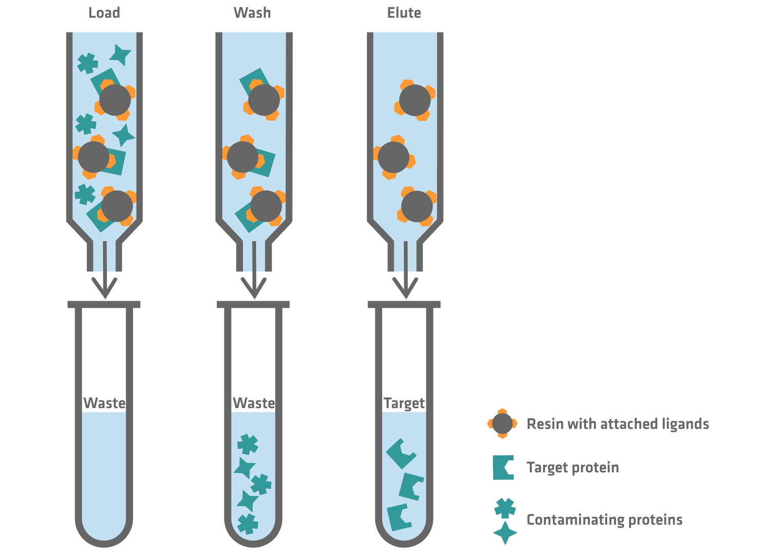
Affinity chromatography (AC) uses columns packed with a resin consisting of tiny beads with attached ligands. When the sample is loaded into the column, the target protein attaches to the ligands on the resin and contaminating proteins can be washed away. In the subsequent elution step, the conditions are changed so that the purified target protein is released from the resin and can be collected.
Ligands used for AC can either be:
- known natural binding partners of the target protein, such as enzymes
- antibodies raised against the target protein, obtained by injecting the target protein into a host and purifying the resulting antibodies produced by its immune system
- or, in the case of recombinant protein purification, tag-based ligands. When producing proteins recombinantly, scientists usually engineer cells to express the protein of interest with an affinity tag, which can then be used for AC purification.
AC is a powerful method, allowing scientists to go from a crude lysate to a high purity sample in a single step, by exploiting a specific binding event rather than a general physical property. However, resins used for AC are more expensive than resins made for other chromatographic methods. In addition, AC only works for proteins that either have known natural binding partners, could be purified for antibody production using other methods beforehand, or have been produced recombinantly.
An alternative to column-based AC is the use of magnetic beads functionalized with the same types of ligands. Instead of packing a column, the magnetic beads are mixed directly with the sample, allowing the target protein to bind the ligand in suspension. A magnet is then applied to the outside of the tube or well to pull the beads – and the bound target protein – out of solution, while the unbound contaminants are removed with the supernatant. After a wash step, the target protein is eluted from the beads in a similar way as in column-based AC.
Ion exchange chromatography
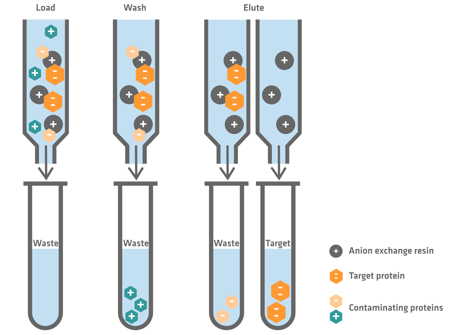
Proteins are either positively or negatively charged. That net charge isn't fixed and depends on the pH of the surrounding solution. Ion exchange chromatography (IEX) exploits this property to separate proteins.
The technique uses a column packed with resin beads that carry a fixed charge of their own. You can either use a cation exchange resin that binds positively charged proteins, or an anion exchange resin that binds negatively charged proteins. The choice between the 2 comes down to a practical question: at a pH the protein can tolerate, is your target protein net positive or net negative?
The separation itself happens in 3 stages.
- The sample is loaded onto the column in a low-salt buffer. Any protein carrying a net charge opposite to that of the resin will bind to it, with proteins carrying a higher net charge generally binding more strongly. Proteins with a very low net charge, or the same net charge as the resin, will remain unbound.
- The column is washed to remove unbound molecules.
- The bound proteins are eluted by gradually increasing the salt concentration in the buffer. Salt ions compete with the proteins for the binding sites on the resin and, as salt concentration rises, they progressively outcompete and displace the bound proteins. Proteins that are bound weakly let go first at low salt concentration, and proteins that are bound more tightly hold on for longer and release only at higher salt concentration. This way, proteins can be separated based on their charge.
IEX is often used to isolate native proteins when affinity purification isn't an option or is too expensive. Moreover, it is ideal for high resolution polishing steps towards the end of a purification process, as its sensitivity to small charge differences allows it to separate the target protein from very close relatives, such as those with different post-translational modifications or conformational changes that alter the protein's surface charge distribution.
Size-exclusion chromatography
Size-exclusion chromatography (SEC), also known as gel filtration, separates proteins according to their size and shape. It uses a column packed with porous beads with a defined pore size; smaller proteins enter the pores and take a longer, more circuitous path through the column, while larger proteins are excluded from the pores and pass through more quickly. As a result, large proteins and protein complexes elute first, followed by progressively smaller molecules.
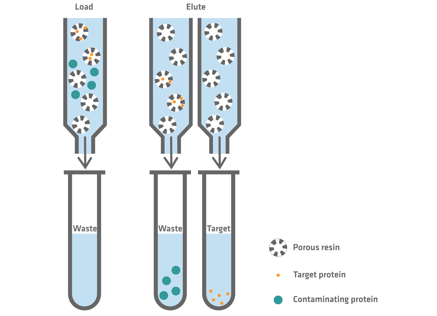
SEC is a gentle purification method as it involves no binding or elution chemistry that could disrupt the protein’s native structure. It is often used as a final polishing step to remove high molecular weight aggregates or low molecular weight degradation products. Moreover, it can be used to transfer a protein sample into a different buffer, e.g. if a sample needs to be desalted after IEX. Small salt molecules from the previously used buffer enter the beads whereas the larger protein molecules quickly pass through the column and elute first, allowing scientists to collect them in a different buffer.
Hydrophobic interaction chromatography
Hydrophobic interaction chromatography (HIC) separates proteins based on the hydrophobicity of their surface. Almost all proteins have at least some hydrophobic character on their surface, but the degree varies from protein to protein, forming the basis for separation.
The sample is run through a column packed with resin carrying mildly hydrophobic ligands. On their own, these wouldn't interact much with proteins in water. The trick is salt: proteins are loaded onto the column in a high-salt buffer. High salt concentrations push water molecules away from hydrophobic surfaces, which forces hydrophobic patches on the protein and resin into contact with each other instead. Elution then works in reverse: the salt concentration is gradually decreased. As salt drops, the hydrophobic interactions weaken, and proteins are released from the column in order of increasing hydrophobicity; the least hydrophobic proteins elute first, the most hydrophobic elute last.
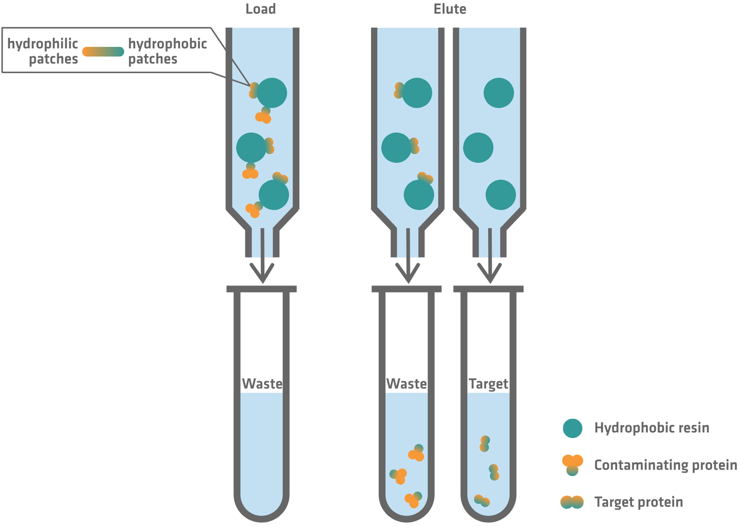
HIC is a convenient next purification step after ammonium sulfate precipitation or IEX, both of which leave the target protein in a high-salt buffer. It is also a comparatively gentle purification method; the interactions involved are typically weaker than ionic or affinity binding, and protein structure and biological activity are preserved. However, not all proteins tolerate exposure to high salt concentrations, and very hydrophobic proteins can bind too tightly and refuse to elute cleanly, making their recovery cumbersome and adding a risk of denaturation.
Dialysis and ultrafiltration
Dialysis and ultrafiltration separate molecules according to size using a semi-permeable membrane. In dialysis, a sample is sealed inside a membrane and placed into a larger volume of buffer. Small molecules, such as salts, diffuse passively through the membrane until equilibrium is reached, while the larger target protein is retained. Ultrafiltration achieves a similar effect faster by applying pressure, typically through centrifugation, to force buffer and small solutes through the membrane, thereby retaining and concentrating the protein.
Both methods are commonly used to exchange buffers, remove small contaminants, and – in the case of ultrafiltration – concentrate a protein sample, rather than as standalone purification techniques. Dialysis is simple and gentle, but time consuming, while ultrafiltration is faster and often used to concentrate dilute protein samples. However, the pressure applied during ultrafiltration can cause aggregation or loss of activity in shear-sensitive proteins.
Protein isolation and purification techniques compared
This table provides a concise comparison of the main protein purification methods discussed above, summarizing the properties they each exploit and identifying where they typically fit into a purification protocol.
| Technique | Basis of separation | Common use |
|---|---|---|
| Centrifugation | Size, density and buoyancy | Initial, crude purification |
| Precipitation | Solubility | Initial, crude purification or concentration |
| AC | Specific ligand binding | Single-step capture of:
|
| IEX | Net surface charge | Alternative to AC to isolate native proteins without binding partner or antibody Polishing step to separate the target protein from very close relatives |
| SEC | Size and shape | Polishing step to remove high molecular weight aggregates or low molecular weight degradation products Buffer exchange if sample needs to be desalted |
| HIC | Surface hydrophobicity | Complement purification step after ammonium sulfate precipitation or IEX |
| Dialysis / ultrafiltration | Molecular size | Buffer exchange and removal of small contaminants Concentration (only ultrafiltration) |
Protein characterization and quantification
Once purification is complete, the resulting sample needs to be analyzed before it can be used in downstream applications. This typically involves answering 4 key questions: is this the correct protein (identity), how pure is it (purity), how much protein is present (concentration) and, if relevant, does it still function as expected (biological activity)?
Identity and purity are often assessed together, most commonly using sodium dodecyl sulfate-polyacrylamide gel electrophoresis (SDS-PAGE). In this method, proteins are separated by size on a gel, allowing researchers to quickly evaluate the sample. A strong band at the expected size suggests the target protein is present, while additional bands indicate contaminants. However, SDS-PAGE alone cannot definitively confirm identity, as different proteins can have similar sizes, and therefore migrate to the same position on the gel. For greater confidence, more specific methods can be used, such as Western blotting, which detects the target protein using antibodies, or mass spectrometry, which can precisely identify proteins and detect modifications or low-level impurities. Analytical SEC is also commonly used to assess sample quality by detecting aggregates or breakdown products.
Protein concentration is most often measured by UV absorbance at 280 nm, a fast and simple method that requires only a small amount of sample. However, other molecules such as nucleic acids can also absorb at this wavelength. When working with less pure samples, dye-based colorimetric assays such as Bradford or bicinchoninic acid (BCA) are therefore preferred, as they are less affected by contaminants. The Bradford assay uses the Coomassie brilliant blue dye that turns more intensely blue when it binds proteins, while the BCA assay is based on proteins reducing copper ions, which subsequently form a purple complex with BCA. In both cases, the color intensity is measured with a spectrophotometer to determine protein concentration.
There are several ways to assess biological activity if necessary. For example, enzymes can be tested using activity assays that measure substrate conversion, while antibodies can be evaluated using binding assays such as ELISAs. This step is important because a protein can appear pure and correctly sized but might still be inactive if it is not properly folded.
As with purification itself, no single method provides a complete picture. Instead, researchers combine several complementary techniques to ensure that the protein is suitable for its intended application.
Common applications of protein purification
Purified proteins drive progress and innovation throughout the entire life sciences sector.
Basic research
In basic research, protein purification helps scientists to understand how proteins work. Purified proteins make it possible to study enzyme activity, protein-protein and protein-nucleic acid interactions, as well as signaling pathways. They are also essential for structural and biophysical studies, where researchers look at how a protein’s shape relates to its function.
Biotechnology
In biotechnology, protein purification is used to produce proteins that serve as tools, reagents and industrial catalysts. Examples include enzymes for food processing, detergents and biofuels, as well as affinity reagents and assay components such as ELISA capture antigens or detection antibodies. These proteins must be purified to defined quality levels so that they behave consistently in manufacturing and analytical workflows.
Drug discovery and biopharmaceuticals
In drug discovery, target proteins need to be purified so that they can be used to test candidate drugs. In the majority of cases, the protein of interest is a membrane protein that is naturally embedded in a lipid bilayer, making the isolation in a stable, functional form especially challenging. Several purification approaches have been developed, e.g. the detergent-free extraction, stabilization and affinity purification in a fully automated 96 well workflow using PlateX MP™ plates from Cube Biotech in combination with the ASSIST PLUS pipetting robot.
In biopharmaceutical production, therapeutic proteins such as monoclonal antibodies or recombinant hormones must be purified to very high standards to meet regulatory requirements and minimize the risk of adverse reactions. In addition, purified proteins are used as antigens in vaccines and as reference standards in diagnostic tests.
Conclusion
Protein purification should be seen as a strategy that involves choosing the right combination of steps to reach the required purity and yield for a given downstream application, while preserving the protein's function if biological activity is important. Centrifugation and precipitation offer fast, inexpensive ways to remove bulk contaminants, chromatographic methods add the resolving power to isolate a target protein to near-homogeneity, and techniques like dialysis and ultrafiltration handle buffer exchange and concentration. Ultimately, efficient protein purification depends on selecting the right sequence of steps, and knowing when the required level of purity has been achieved.
Do you have a purification challenge or a technique you rely on that we didn't cover? Let us know in the comments below.
Ask our expert. Leave a comment!
Write us if you have any questions regarding the blog article.